Click here to close
Hello! We notice that you are using Internet Explorer, which is not supported by Xenbase and may cause the site to display incorrectly.
We suggest using a current version of Chrome,
FireFox, or Safari.
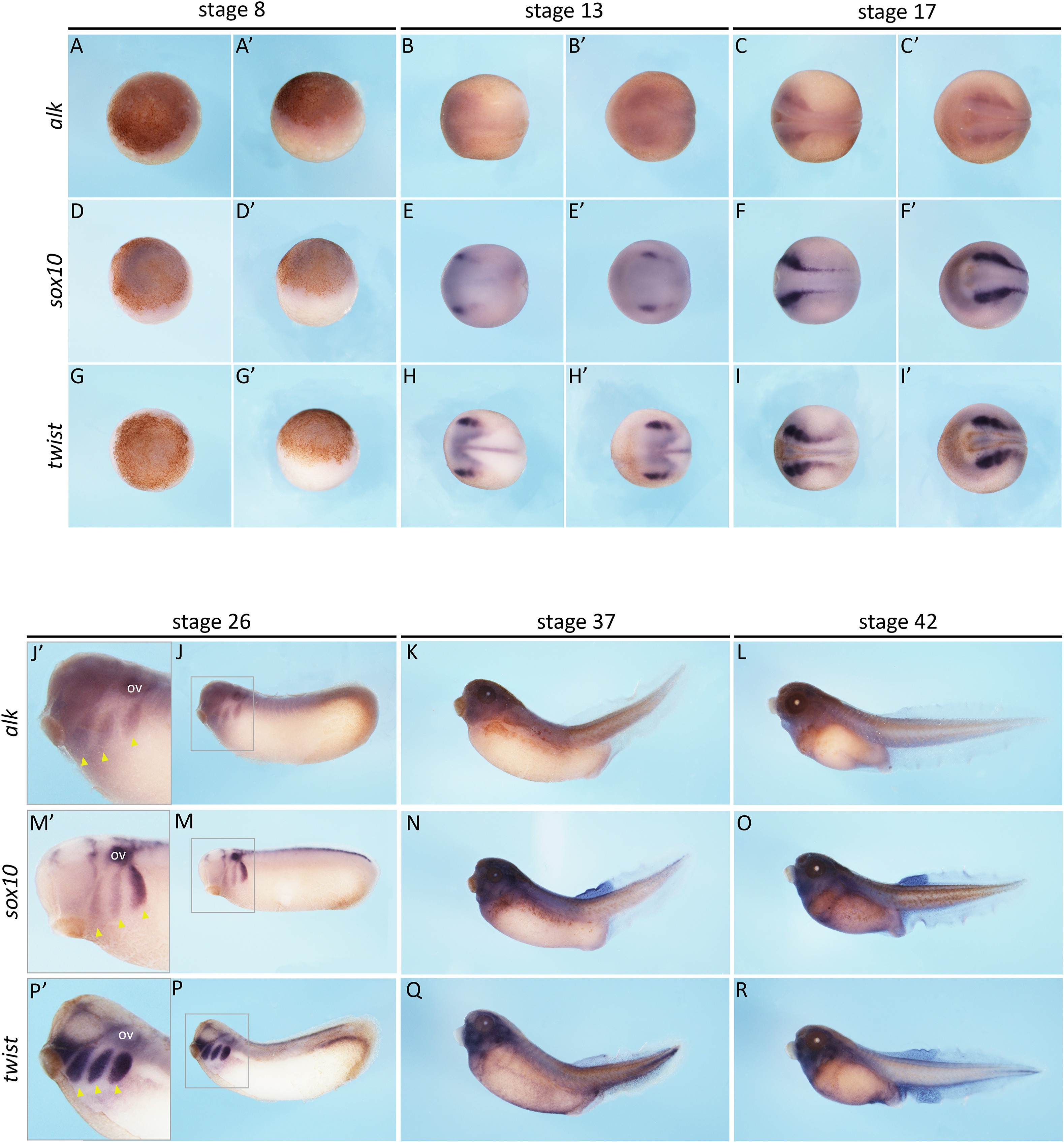
Fig. 2. alk expression pattern in Xenopus laevis. mRNA in situ hybridisation for alk and neural crest markers, sox10 and twist, in Xenopus laevis embryos at stages 8, 13, 17, 26, 37 and 42 (Nieuwkoop and Faber, 1994). Animal (A, D and G) and lateral (Aâ², Dâ² and Gâ²) view of stage 8 embryos (alk n = 11, sox10 n = 5, twist n = 4). Dorsal (B, E and H) and anterior (Bâ², Eâ² and Hâ²) view of stage 13 embryos (alk n = 12, sox10 n = 4, twist n = 2). Dorsal (C, F and I) and anterior (Câ², Fâ² and Iâ²) view of stage 17 embryos (alk n = 7, sox10 n = 4, twist n = 4). Lateral view (J, M and P) of stage 26 embryos and zoomed insets (grey rectagles) of embryos heads (Jâ², Mâ² and Pâ²), showing otic vesicles (OV) and neural crest streams (yellow arrowheads) (alk n = 8, sox10 n = 7, twist n = 5). Lateral view (K, N and Q) of stage 37 embryos (alk n = 10, sox10 n = 7, twist n = 5) and stage 42 embryos. Lateral view (L, O and R) of stage 42 embryos (alk n = 11, sox10 n = 5, twist n = 7).