XB-IMG-212129
Xenbase Image ID: 212129
|
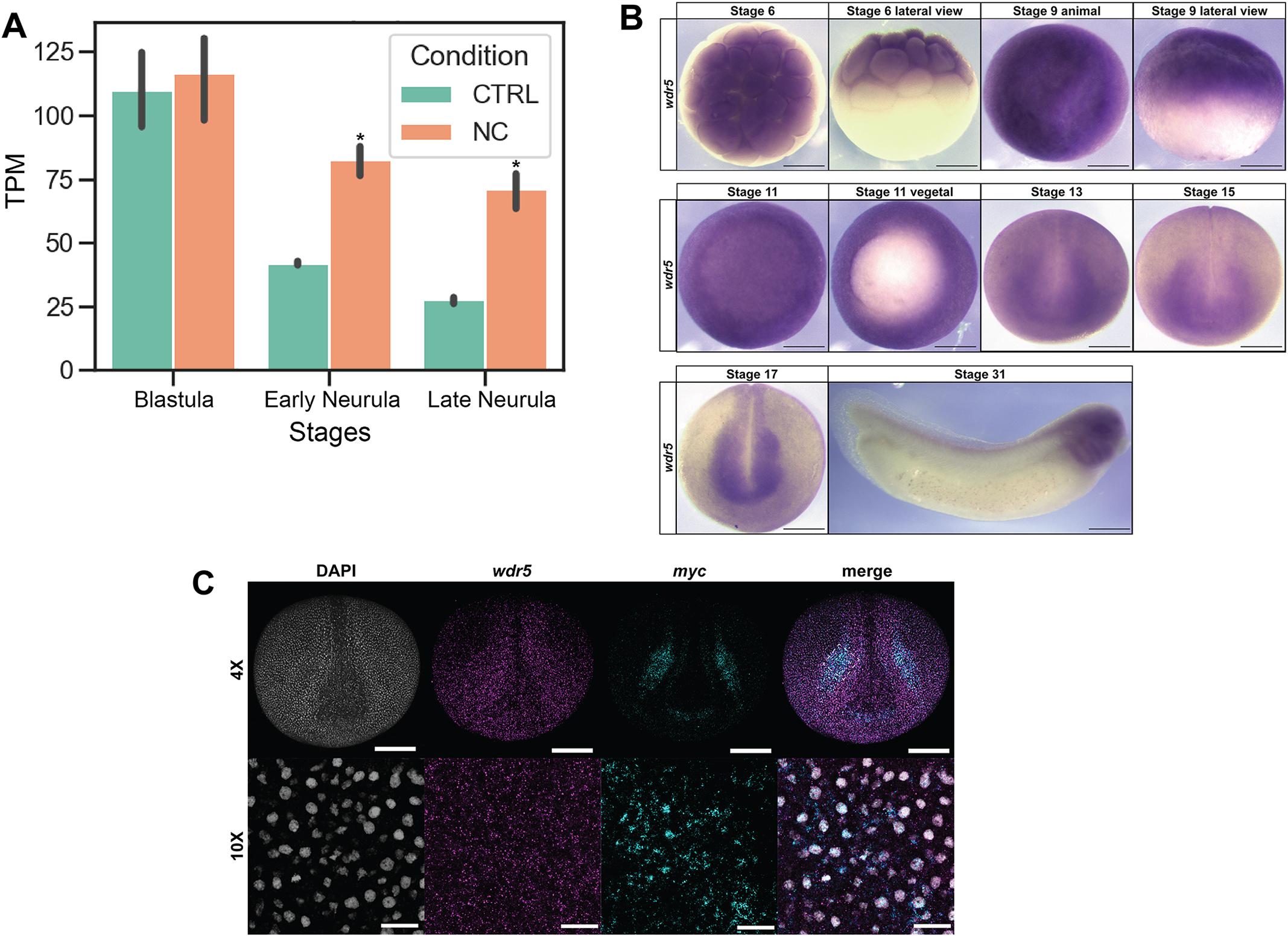
Fig. 1. Characterization of wdr5 expression. (A) Analysis of previously published RNA-Seq data (York et al., 2024) shows wdr5 transcripts are abundant in the animal pole cells of the blastula stages and differentially enriched in neural crest-induced animal caps at early and late neurula stages (*P<0.05). (B) Spatial and temporal expression of wdr5 shows that wdr5 transcripts are maternally provided, enriched in blastula stem cells and retained broadly throughout the neuroectoderm into developing neural crest stem cells and neural crest derivatives. (C) Fluorescent in situ hybridization chain reaction confirms wdr5 expression in the neural crest that colocalizes with myc a canonical neural crest and pluripotency factor. CTRL, control; NC, neural crest; TPM, transcripts per million. Scale bars: 250[micrometers] in B and C (10 images); 100[micrometers] in C (4 images). Image published in: Compton K et al. (2026) © 2026. Creative Commons Attribution license Larger Image Printer Friendly View |